Computer graphics A set of devices, methods, and procedures to visualize and manipulate visual object representations in a virtual environment.
Computed tomography (CT) X-ray-based imaging method that permits reconstruction of cross-sectional images of patients and other objects from radiographic projections. geometric morphometrics (GM) Methods of quantification of organismic form that integrate real-space geometric properties of the morphology of organisms into multivariate analysis. virtual reality (VR) A computer-based environment in which a user interacts with geometric object representations using input and output devices that provide perceptual equivalence to physical reality.
Real virtuality (RV) An environment in which a user interacts with physical models of three-dimensional objects generated or modified by computer-assisted procedures. rapid prototyping (RP) Technologies that transform objects from virtual reality back to the physical world.
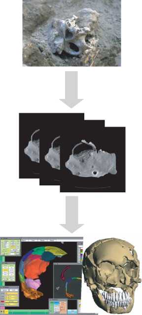
Computer-assisted palaeoanthropology (CAP) is a new discipline that emerged with the advent of novel techniques in the fields of biomedical imaging, computer graphics, rapid prototyping, and geometric-morphometric analysis. The major reason to bundle these disciplines into a coherent transdisciplinary approach is to tackle a central problem of palaeoanthropology, namely the scarcity of the fossil record and the incompleteness of most specimens. In order to gain a maximum of information from the scant but precious material evidence, the primary aim of CAP (see Paleoanthropology) is to enhance the investigative power while minimizing the invasiveness of methods used for fossil data acquisition, preparation, reconstruction, anatomic diagnosis, and comparative morphometric analysis. The basic concept of CAP is to enable the user to perform these tasks in a ‘virtual reality’ setting (Figure 1).
The first step in a typical CAP workflow is the acquisition of three-dimensional (3D) data from a fossil specimen - which may still be embedded in sediment - with the technique of ‘computed tomography’ (CT). The development of CT in the 1970s marked an important step toward the improvement of noninvasive medical diagnostics, as it permits acquisition of serial cross-sectional image data from patients. CT was quickly adopted for ‘fossil diagnostics’, giving insights into internal anatomic features of fossil specimens, and into regions still covered by sediment. Parallel to these
Fossil in matrix
Volume data acquisition
Virtual preparation and reconstruction
/IV
Physical
Replicas
Morphometric
Analysis
Morphogenetic
Modeling
Figure 1 CAP consists of three main steps. Digital volume data of hominid fossils sfill embedded in sediment are acquired with CT. All subsequent steps are performed noninvasively through interactive computer graphics tools. Virtual preparation is performed with data segmentation tools. Segmented data are transformed subsequently into 3D object representations, which can be manipulated interactively on the computer screen to perform reconstructive tasks. Finalized virtual fossils can be transformed into physical models with RP technology. Virtual sampling of two - and three-dimensional measurements permits extensive comparative morphometric analyses. Computer models of processes of taphonomy, evolution, and development lead to a better understanding of the causes underlying fossil form variation.
Developments the rapid increase of computer power and advancements in the area of image processing and ‘computer graphics’ opened up new fields in the visualization and interactive manipulation of 3D objects in a virtual environment. In CAP, these techniques build the basis for virtual fossil reconstruction. Applying 3D data segmentation procedures to volumetric CT data sets, it is possible to free a fossil fragment embedded in sediment with a ‘computer chisel’, and to extract internal anatomical structures such as the labyrinthine cavities of the inner ear. After data segmentation, a geometric description and a graphical representation of the fossil fragments are created through 3D reconstruction procedures.
These fragments are then recomposed in virtual reality. This procedure is analogous to the assembly of a 3D puzzle in which important pieces are deteriorated or have been lost. The primary aim of virtual reconstruction is to reverse the fossilization process in order to infer the morphology of the specimen at the
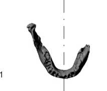
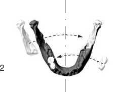
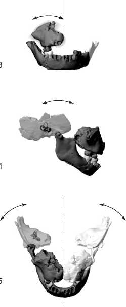
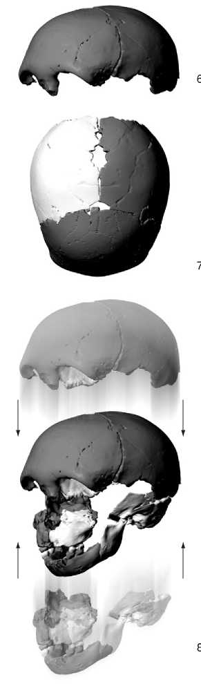
Figure 2 Virtual reconstruction of a fragmentary Neanderthal child skull from the site of Devil’s Tower, Gibraltar. Only five original fragments are preserved (dark gray): parts of the mandible, the right maxillary and temporal bones, the frontal bone and associated left parietal bone. Virtual reconstruction proceeds in eight steps. 1: orientation of the fragmentary mandible along the virtual midplane of the skull; 2: mirror-image completion (white) of missing left mandibular ramus and right milk molars; 3: establishment of dental occlusion between mandibular and maxillary dentition; orientation of maxilla relative to midplane; 4: articulation of the right temporal bone with the maxilla and orientation according to the anatomy of the inner ear; 5: mirror image completion of missing parts on the left side; 6: articulation of frontal bone and left parietal bone; 7: mirror image completion of missing right parietal bone; and 8: docking of independent reconstructions of the face/cranial base and cranial vault.
Time of its death (Figure 2). This involves correction of distortions that occurred during fossilization, reestablishment of anatomic connections between preserved parts, and completion of missing parts by mirror-imaging preserved antimeres. During this process, each reconstructive step follows predefined anatomic criteria. This prevents a reconstructive bias toward preconceived morphologies, ensures reproducible results, and makes the entire procedure accessible to other researchers. Additionally, the reliability of the process as a whole can be evaluated by creating a range of potential reconstructions.
Virtual reconstruction is an iterative process in its essence, in which reconstructive attempts can be gradually refined without physically dismantling earlier versions and exposing the original specimens to further damage. A major advantage during this process is the absence of physical forces such as gravitation. Each piece of the fossil puzzle can be fixed in virtual anatomic space without the need for glue, plaster, or stabilizing elements.
Handling fossil fragments in virtual reality instead of physical reality is of immediate benefit. Whereas physical preparation, manipulation, and reconstruction are highly invasive and potentially destructive processes, the analogous computer-assisted procedures are completely noninvasive. Additionally, CT volumetric data provide unlimited access to all external and internal anatomic structures from an infinite number of views.
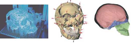
The finalized virtual reconstruction of a fossil represents the basis for various further analyses and procedures: virtual fossils can be used as a guide during physical preparation, transformed into physical hard copies, and submitted to comparative morphometric analysis.
Transforming a fossil reconstruction from virtual reality into ‘real virtuality’, that is, into a physical hardcopy, is typically achieved with rapid prototyping (RP) technologies. Awidely used method is stereolithography, where, analogous to the construction of topographic models from piles of cardboard layers, the object is constructed from automated superpositioning of cross-sectional layers. In this manner, it is possible to build models containing arbitrary geometric complexity, including cavities, in a single production step. Stereolithographic replicas of fossil specimens - before and after virtual reconstruction - represent a valuable noninvasive and secure alternative to classic mold-and-cast techniques. RP replicas convey touch-and-feel information and are thus ideal for teaching purposes and for interactive museum exhibits. Similarly, RP models drawn at different stages of virtual reconstruction can be used to monitor spatially complex matching tasks.
Comparative morphometric analyses of reconstructed fossil specimens represent an essential part of CAP and range from functional and biomechanical analysis to the analysis of patterns of evolutionary diversification and developmental differentiation.
Because virtual fossils are graphical objects, they provide an ideal basis for exploration of a wide variety of one-, two-, and three-dimensional measurements. By supplementing the classic repertoire of rulers and calipers with virtual morphometric tools, spatial position of anatomic landmarks, areas of muscular attachment, paranasal sinus volumes, surface curvatures, bone thickness distribution, moments of inertia, strain distribution, and so on can be measured. The new field of ‘geometric morphometrics’ (GM) is of special relevance for CAP, because GM methods integrate real-space geometric properties of 3D biological forms into multivariate analyses. Using special-purpose computer graphics tools, it is possible to explore complex patterns of shape variability in multivariate space and to visualize them as 3D patterns of shape change in virtual organismic forms. These methods have proved instrumental to identify differences and commonalities between patterns of growth in fossil and extant hominoid species. CAP also comprises computer simulations of form variation in fossils, be it to test hypotheses regarding taph-onomic deformation of a given fossil specimen, or to test more general hypotheses about how empirical patterns of form variability are related to underlying evolutionary and developmental processes.
The possibility of archiving and retrieving virtual fossil specimens opens new avenues of comparative analysis. Making use of an extended database of digital fossils and the virtual desktop, it becomes possible to inspect fossil specimens that are literally separated by continents in side-by-side comparisons and to derive new measurement series for testing hypotheses without requiring re-examination of the original specimens. However, virtual hominid fossils also raise questions of copyright and access regulations that still need to be resolved. Hominid specimens often represent national treasures, and many of the most valuable specimens come from countries where access to scientific knowledge, especially in its digital form, is severely hampered by infrastructural, educational, and political hurdles. Problems arising from the resulting unidirectional knowledge transfer are similar to problems associated with ‘gene trading’.
See also: Anthropological Archaeology; Paleoanthropology.
 World History