Bone disorders arising from congenital conditions, those present at birth or that develop in the course of growth and development, can also be observed in skeletal remains. These may be due either to trauma during birth or to an underlying genetic predisposition, sometimes with a link to environmental pollutants and maternal health and nutrition. Palaeo-pathological studies of congenital conditions have tended to concentrate on those producing a developmental absence of an entire anatomical structure or a part thereof, examples being spina bifida occulta, an open vertebral canal, or mid-line defects such as cleft palate, or skeletal dysplasia, such as seen in club-foot deformity (talipes equinovarus) that develops in utero, or muscular torticollis (wryneck deformity) and hip dysplasia from damage to the nerves and/or musculature from traction injuries (i. e., pulling) during a difficult birth (Figure 17).
Many congenital conditions are identified based on the changes they bring not only to morphology, but also to shape, size, and proportion. Among the most commonly identified in the archaeological record is achondroplasia or dwarfism, from a mutation of the FGFR3 gene on chromosome 4, which alters the relative proportions of the limbs with respect to the axial skeleton, producing short stature. Down’s syndrome or trisomy 21, linked to non-disjunction of chromosome 21 in the ovum inherited from the mother, has also been identified due to the effect that this disorder has on cranial shape and dimensions. Since many congenital syndromes influence cranial development, a similar approach using craniometrics and larger population samples will increase what is, at present, an under-explored area of enquiry. Since many of these conditions produce mental impairment (e. g., a lack of or reduction in normal function), their identification in the archaeological record permits insight into the social circumstances that contribute not only to their occurrence but also to the survival and perceptions of people with identified mental disability from their burial treatment. Because these types of
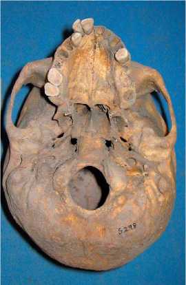
Figure 17 The cranial asymmetry and twisting of the cranial base seen in muscular torticollis or wryneck deformity in a Medieval adult female from St. Helen’s-on-the-Walls, York, from the collections of the Yorkshire Museum. The left mastoid process is atrophied, the occipital flattened on the right, with slight bulge on left, and the right parietal is larger than that of the left side. Courtesy of Rebecca Storm, Biological Anthropology Research Centre (BARC), University of Bradford, from the collections of the Yorkshire Museum. From: Storm RA (forthcoming) Human Skeletal Asymmetry: A Biometrical Study of Fluctuating Asymmetry to Assess Health and Social Status of English Populations from the 7th to the 19th Centuries. Unpublished PhD Thesis, University of Bradford.
Conditions often affect both appearance and behavior, they are ideal candidates for the application of the biocultural approach that would permit assessment of social reactions to the impaired.
 World History